Contents
Overview
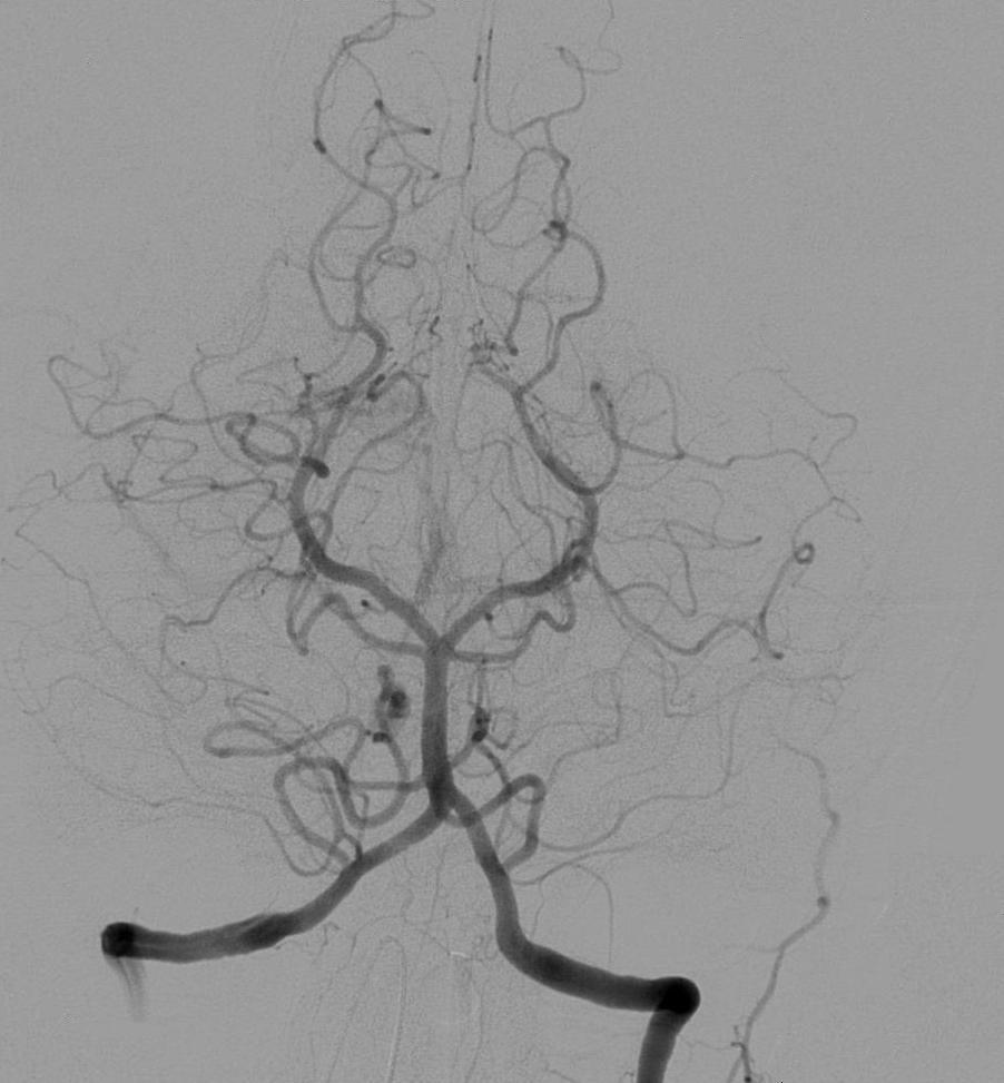
Angiography is a sophisticated medical imaging technique that illuminates the intricate network of blood vessels within the human body. It functions by introducing a radio-opaque contrast agent, which acts like a dye under X-ray, allowing physicians to visualize the flow and structure of blood vessels in real-time. This diagnostic tool is crucial for identifying blockages, aneurysms, malformations, and other vascular abnormalities that might otherwise remain hidden. While traditionally reliant on X-ray fluoroscopy, advancements like magnetic resonance angiography (MRA) now offer contrast-free visualization, expanding its diagnostic reach and patient comfort. The resulting images, known as angiograms, are indispensable for guiding minimally invasive treatments and complex surgical interventions, fundamentally shaping cardiovascular and neurological care since its inception.
🎵 Origins & History
The genesis of angiography traces back to the early 20th century, a period brimming with innovation in medical imaging. The development of safer, water-soluble contrast agents in the 1930s and 1940s significantly improved the safety and widespread adoption of the technique, moving it from experimental curiosity to a clinical staple.
⚙️ How It Works
At its core, angiography is a visual narrative of the circulatory system. The process typically begins with the insertion of a catheter, a thin, flexible tube, into a major artery or vein, often in the groin or arm. Guided by real-time fluoroscopy (a type of X-ray imaging), this catheter is advanced to the specific blood vessel or organ of interest. A contrast agent, usually iodine-based, is then injected through the catheter. This agent, which absorbs X-rays more effectively than surrounding tissues, makes the blood vessels appear bright white on the X-ray images. A series of rapid X-ray exposures captures the contrast agent's movement through the vessels, creating a dynamic map of blood flow. Newer techniques like Magnetic Resonance Angiography (MRA) bypass the need for ionizing radiation and contrast agents by utilizing magnetic fields and radio waves to map blood flow.
📊 Key Facts & Numbers
The scale of angiography's impact is staggering. The market for angiography devices and consumables is projected to exceed $10 billion by 2025, underscoring its economic importance. Angiography boasts a sensitivity rate of over 95% for detecting significant arterial stenoses (narrowing) above 50%. The average procedure time for a diagnostic coronary angiogram is typically between 30 to 60 minutes, with complications occurring in less than 1% of cases when performed by experienced operators at high-volume centers.
👥 Key People & Organizations
Pioneers like Egas Moniz and Raymond W. Whittington are foundational figures in angiography's history. In contemporary practice, interventional radiologists and interventional cardiologists are the primary practitioners. Organizations such as the Society of Interventional Radiology (SIR) and the American College of Cardiology (ACC) play crucial roles in setting standards, conducting research, and educating professionals. Companies like GE Healthcare, Siemens Healthineers, and Philips Healthcare are major innovators, developing advanced angiography systems and contrast agents that push the boundaries of diagnostic and therapeutic capabilities.
🌍 Cultural Impact & Influence
Angiography has profoundly reshaped patient outcomes and medical understanding. Its ability to visualize previously unseen vascular pathologies has led to the development of minimally invasive treatments, reducing the need for open surgery. Procedures like angioplasty and stent placement, guided by angiography, have become standard for treating conditions like coronary artery disease and peripheral artery disease. The visual data generated by angiograms has also fueled countless research studies, advancing our knowledge of vascular biology and disease progression. Its influence extends beyond clinical practice, appearing in medical dramas and documentaries, raising public awareness of cardiovascular health and interventional medicine.
⚡ Current State & Latest Developments
The field of angiography is in constant flux, driven by technological advancements and a push for less invasive diagnostics. Cone-beam CT angiography is gaining traction for its ability to provide high-resolution 3D images with faster acquisition times. Innovations in artificial intelligence are being integrated to automate image analysis, detect subtle abnormalities, and predict procedural risks. Furthermore, the development of novel, lower-osmolality and even non-iodine-based contrast agents aims to further reduce patient risks and allergic reactions. The trend is clearly towards more precise, faster, and safer vascular imaging, with a growing emphasis on integrating imaging data with other patient information for personalized treatment planning.
🤔 Controversies & Debates
Despite its immense utility, angiography is not without its controversies. The use of ionizing radiation and iodinated contrast agents carries inherent risks, including radiation exposure, contrast-induced nephropathy (kidney damage), and allergic reactions, though these are generally low in experienced hands. Debates persist regarding the optimal timing and necessity of angiography for certain conditions, particularly in the era of advanced non-invasive imaging like CT angiography and MRA. The cost-effectiveness of routine angiography versus less invasive alternatives is also a recurring discussion point within healthcare systems. Furthermore, the ethical considerations surrounding incidental findings and the potential for over-diagnosis remain subjects of ongoing professional dialogue.
🔮 Future Outlook & Predictions
The future of angiography points towards even greater integration with interventional procedures and a move towards 'hybrid' imaging suites. We can anticipate further refinement of AI algorithms for real-time image guidance and automated analysis, potentially reducing procedure times and improving accuracy. The development of 'smart' contrast agents that can provide functional information beyond just anatomical visualization is on the horizon. Furthermore, advancements in catheter technology, including micro-robots and improved navigation systems, will enable access to smaller and more complex vascular territories. The ultimate goal is to make vascular imaging and intervention as seamless and non-invasive as possible, transforming it from a diagnostic tool into a fully integrated therapeutic platform.
💡 Practical Applications
Angiography's practical applications are vast and critical across numerous medical specialties. In cardiology, coronary angiography is the gold standard for diagnosing coronary artery disease and guiding interventions like percutaneous coronary intervention (PCI). Neurology utilizes cerebral angiography to detect strokes, aneurysms, and arteriovenous malformations (AVMs). In vascular surgery, it's essential for planning procedures on arteries in the limbs, abdomen, and neck, treating conditions like peripheral artery disease and aortic aneurysms. Angiography also plays a role in oncology, helping to map tumor vascularity for targeted therapies and embolization, and in nephrology for diagnosing and treating kidney-related vascular issues. Interventional radiologists use it extensively for embolization procedures to stop bleeding or treat tumors.
Key Facts
- Category
- technology
- Type
- topic